Microarray Printing Buffers Details Page
information, details and purchase
Microarray Printing Buffers
Protein Printing Buffer Plus to Enhance Protein Microarray Control Sample Manufacturing
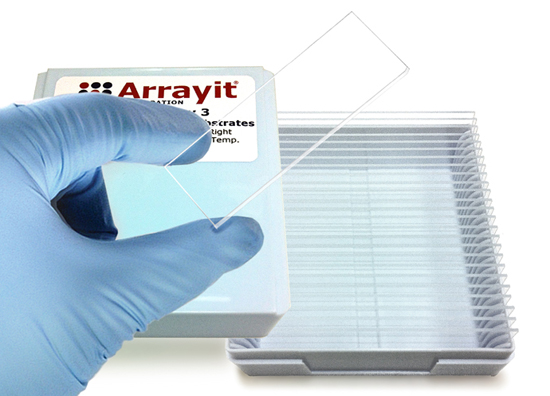
Arrayit advanced formulation for peptide and protein microarray manufacturing that enhances the precision and printing quality of protein dilution series controls such as IgG, anti-IgG and other sample types essential for microarray testing. Use Protein Printing Buffer Plus on SuperEpoxy 2 and 3 glass substrate slide surfaces when printing dilution series of control proteins. This buffer is delivered at a 50 ml of 2X solution.
``````````Additional Information
Table of Contents
- Introduction
- Quality Control
- Product Description
- Technical Note
- Technical Assistance
- Short Protocol
- Complete Protocol
- Recommended Equipment
- Troubleshooting Tips
- Ordering Information
- Warranty
Introduction
Congratulations on taking a big step towards improving the economies of scale, quality and speed of your proteomics research. This booklet contains a complete set of protocols outlining the steps and principles needed to use Arrayit Protein Printing Buffer.
Quality Control
Arrayit assures the performance of this product. The finest scientific research went into the development of this product. Rigorous quality control monitoring on a lot-by-lot basis guarantees that the ingredients conform to the highest industry standards.
Product Description
The Arrayit Protein Printing Buffer Plus (PRP) is an advanced buffering system containing a proprietary mixture of ionic and polymeric materials. A specially formulated buffer for microarray manufacturing of dilution series control proteins on SuperEpoxy 2 and 3 Microarray Surface Chemistry. Not for use on membrane surfaces.
Users will appreciate the following features of Protein Printing Buffer Plus (PRP):
- Supports Arrayit’s most widely used printing technology
- Print proteins, enzymes, antibodies, receptors, antigens, and peptides
- Washes away during blocking and processing
- Manufacture microarrays of pure proteins, recombinant proteins and cellular extracts
- Stabilizes protein samples and prevents denaturation
- Provides uniform feature size and coupling density accross lowering concentrations
- Protects printed samples from environmental damage
- Slows sample evaporation within the source microplates
- Minimizes sample drying and crystallization on substrates and pins
- Washes away easily, leaving pure bound protein molecules
- Improves deposition uniformity which facilitates data analysis
- Arrives pre-mixed as a 2X solution, sterile and protease-free
- Sufficient to print 50 million protein features (5,000 10K protein arrays)

Figure 2. Ten Fold Dilution series of Human IgG starting at 1 µg/µl Printed in Protein Printing Buffer Plus left to right top down. Microarrays manufactured using a NanoPrint Microarrayer, Professional Printhead and 946MP3 Microarray Printing Pins. SuperEpoxy2 Microarray Substrates, Blocked wtih BlockIt Plus, reacted with Anti-Human IgG Conjugated to Cy3 using diluted 1 to 2000 in Protein Microarray Reaction Buffer using an RC1x24 Microarray Reaction Cassette. Microarray Scan at 5 um resolution using an Innoscan 710 Microarray Scanner.
Technical Note
It may be necessary to add protease or phosphatase inhibitors to the Protein Printing Buffer to increase the stability of certain proteins or protein extracts. Protein stability can also be enhanced if microarray manufacture is performed at 4°C. The SpotBot and NanoPrint Microarrayers can be operated in a cold room. A special “cold platen” is also available for these machines if a 4°C printing temperature is required in an ambient laboratory environment. Please contact arrayit@arrayit.com for technical details.
Technical Assistance
Please contact us if you have any comments, suggestions, or if you need technical assistance. By electronic mail: arrayit@arrayit.com (under the subject heading, please type, “Technical assistance”). By telephone: (408) 744-1331, Monday–Friday, PST 9:00am - 4:30pm. Please remember that we want to hear about your successes!
Short Protocol (Steps 1-10)
1. Obtain 0.2-1.0 µg/µl protein samples.
2. Transfer 4.0 µl per well of each protein sample into 96- or 384-well microplates.
3. Add 4.0 µl per well of 2X Arrayit Protein Printing Buffer (PPP).
4. Mix the samples by pipetting up and down 10 times.
5. Print protein samples onto SuperEpoxy 2 or 3 .
6. Use BlockIt Plus to block and process the printed protein microarrays.
7. React the processed microarrays with fluorescent samples.
8. Wash the microarrays to remove unreacted fluorescent material.
9. Scan the microarray to produce a fluorescent image.
10. Quantitate and model the fluorescent data.
Complete Protocol (Steps 1-10)
1. Obtain 0.2-1.0 µg/µl protein samples and determine the desired dilution series. Phosphate buffered saline (PBS) at 1X concentration works very well for most proteins. Protein samples should be free of aggregates and particulates that can clog printing devices and impair attachment to the microarray substrate. Aggregates and particulates can be removed by centrifugation or filtration. A 50kD protein at 1 µg/µl concentration has a concentration of 20 µM. At 30% coupling efficiency, a 20 µM protein will produce a target density of 1011 proteins per mm2 of substrate.
2. Transfer 4.0 µl per well of each protein sample into 96- or 384-well microplates. Transfer can be performed manually by pipette or with a liquid handling robot. Certains proteins are fragile and protein samples should be handled with care to avoid damaging the structure and function of proteins.
3. Add 4.0 µl per well of 2X Arrayit Protein Printing Buffer (PRP). This will give a final concentration of 1X Protein Printing Buffer and a final protein concentration of 0.1-0.5 µg/µl. Certain proteins or protein extracts are more stable at 4°C. Keeping the proteins samples cool may improve stability. Stability can also be improved in some cases by the addition or protease and phosphatase inhibitors or by the use of a SpotBot or NanoPrint Microarrayer equipped with a cooled platen.
4. Mix the samples by pipetting up and down 10 times. This step ensures that the protein samples mix thoroughly with the 2X Protein Printing Buffer Plus. Failure to mix the samples thoroughly will produce poor quality microarrays.
5. Print dilution series onto SuperEpoxy 2 or 3 Microarray Substrates. The SuperEpoxy surface couples proteins more readily than other surfaces owing to the increased reactivity of the epoxide groups. Another advantage of SuperEpoxy is that coupling can occur in humid and conditions as may be required to maintain protein structure and function.
6. Use BlockIt Plus blocking buffer and process the printed protein microarrays. Once the printing process is complete, wash the printed microarrays Protein Microarray Wash Buffer to remove unbound protein molecules and components of the printing buffer. Protein binding to the SuperEpoxy 2 and 3 surface is extremely stable and the microarrays can be washed, blocked and reacted without sufficient loss of coupled protein. Allow microarrays to dry overnight to maximize binding. A good blocking protocol involves a 1 hour room temperature to overnight incubation at 4 degrees C in 1X BlockIt Plus, using a High Throughput Wash Station or petri dish. Blocking can be performed with or without agitation. The BlockIt Plus blocking step will inactivate unreacted epoxy groups and prevent background noise. After blocking, wash the microarrays in Protein Microarray Wash Buffer follow by a short few second rinse in Protein Microarray Rinse Buffer. Use High Throughput Wash Station, petri dish or similar device to get gentle agitation for good washing.
7. React the processed microarrays with fluorescent samples. Processed microarrays containing coupled target proteins can be reacted with fluorescent samples to study protein-protein interactions. Binding reactions can be performed using Protein Microarray Reaction Buffer. Fluorescent samples can be incubated as a droplet over the printed microarray, underneath a cover slip, or in a microfluidics chamber. Consider using AHC A 60-minute incubation at room temperature is usually sufficient to obtain strong binding and intense fluorescent signals (see Fig. 1). A Hybridization Cassette can be used to prevent sample evaporation during prolonged binding reactions.
8. Wash the microarrays to remove unreacted fluorescent material. Once binding between the bound target proteins and the fluorescent protein probe molecules is complete, wash the microarray to remove the unbound material. Washes can be performed three times for 5 min each at room temperature in 1X PBS. After the wash procedure, excess buffer should be removed from the surface by tapping or by centrifugation with a Microarray High-Speed Centrifuge.
9. Scan the microarray to produce a fluorescent image. The fluorescent microarray can be scanned or imaging using any of a number of high quality commercial detection instruments, but for best results use Innoscan Microarray Scanners. Instrument settings can be adjusted to optimize the imagine acquistion process.
10. Quantitate and model the fluorescent data using Mapix or other microarray quantification software.
Recommended Equipment
NanoPrint 2 Microarrayers
946 Spotting Device
Stealth Micro Spotting Device
SpotBot® 4 Personal Microarrayers
SuperEpoxy 3 Microarray Substrates
Protein Printing Buffer
BlockIt Blocking Buffer
High Throughput Wash Station
Microarray High-Speed Centrifuge
Microarray Hybridization Cassette
Troubleshooting Tips
Poor printing quality:
- Incomplete mixing of protein samples in Protein Printing Buffer Plus
- Poor printing environment (50% humidity and 25°C recommended).
- Check wash dry station of microarrayer to make sure it is functioning
- Damaged microarary pritning pins
- Not using SuperEpoxy 2 or 3 Surface Chemistry
Poor protein coupling:
- Poor surface chemistry (Use SuperEpoxy 2 or 3)
- Inhibitor in protein sample (suspend in 1X PBS and then 1X PRP)
Weak fluorescent signals:
- Fluorescent protein probe mixture binds poorly to protein targets
- Probe labeling inefficient
- Washes too harsh
*International pricing may vary as much as 30% (or more depending on country) due to import duties, stocking fees and technical support.
*To order ArrayIt® Brand Products: call 408-744-1331, fax 408-744-1711 or click on the purchase button to proceed directly to the purchase page.
Storage Conditions
Arrayit’s ArrayIt® brand Fluorescent Probe Purification Kits should be stored dry at room temperature (20-25°C). The kits perform well across a wide range of ambient temperatures and relative humidity. The kits are nuclease-free, sterile, and have a shelf life of one-year from the date of purchase.
Warranty
ArrayIt® brand products have been scientifically developed and are sold for research purposes. Extreme care and exact attention should be practiced in the use of the materials described herein. All ArrayIt® brand products are subject to extensive quality control and are guaranteed to perform as described when used properly. Any problems with our ArrayIt® brand product should be reported to Arrayit immediately. Arrayit’s liability is limited to the replacement of the product, or a full refund. Any misuse of this product including deviations from our protocols is the full responsibility of the user, and Arrayit makes no guarantees under these circumstances.


