Protein
Data Sheet
![]() Shop this product in our online store
Shop this product in our online store
Products - Microarray Substrates & Slides - Protein A/G Treated Glass Substrate Slides

ArrayIt® Protein A/G Microarray Substrates contain a highly uniform mono layer of Recombinant Protein A/G over the surface of the substrate for microarray applications. The surface is designed to bind human immunoglobulin proteins IgG1, IgG2, IgG3, IgG4 and all types of total IgG. The open platform microscope slide size format format makes this product compatible with all standard microarrayer printers, scanners and other microscope slide processing and detection equipment.
Table of Contents
- Introduction
- Quality Control
- Product Description
- Protocol Protein Microarray
- Technical Specifications
- Technical Assistance
- Recommended Equipment
- Ordering Information
- Storage Conditions
- Warranty
Introduction
Congratulations on taking a big step towards improving the affordability, quality and speed of your genomics, biomedical, pharmaceutical and agricultural research. This booklet contains all the information required to take full advantage of ArrayIt® Protein A/G Microarray Substrates.
Quality Control
Arrayit takes every measure to assure the quality of our ArrayIt® Brand Super Microarray Substrates. The finest microarray biochip cleanroom research was used to develop these products. Rigorous quality control monitoring on a substrate-by-substrate basis guarantees that each these products conforms to the highest industry standards.
Product Description
This surface starts with ArrayIt SuperClean glass then it is activated with Protein A/G. Gene fusion of the Fc-binding domains of Protein A and Protein G has resulted in production of a structurally and functionally chimeric protein with broader binding than either Protein A or Protein G alone. During fusion, the Protein G gene sequence coding for the serum albumin-binding site is eliminated. This protein is expressed in E.Coli and secreted into the surrounding medium during fermentation. The product obtained is consistent in quality and yield because the bacterial host is engineered to be deficient in major proteolytic activities. Binding is less pH-dependent than either Protein A or Protein G alone, occurring well at pH 5-8. The extended Fc-binding properties of Protein A/G make it a popular tool in the investigation and purification of immunoglobulins. Protein A/G binds to all human IgG subclasses, IgA, IgE, IgM and to a lesser extent IgD; however, it does not bind mouse IgA, IgM or murine serum albumin. Protein A/G is an excellent tool for purification and detection of mouse monclonal antibodies from IgG subclasses without interference from these other serum proteins. Individual subclasses of mouse monoclonals are most likely to have stronger affinity to this chimeric protein than to either Protein A or Protein G. The Protein A/G on the substrate is covalently bound to the surface and will not flake, crack or peel off the slide. SuperClean substrate as the foundation for this product is a highly polished glass surface with an optical smoothness tolerance of 50 angstroms over the entire 25 mm x 76 mm surface area for optimum homogeneity; this new surface sets the highest quality standard for Protein A/G-activated substrates for microarray applications. These substrates are packed in white slide boxes, vacuum packed and should be stored at 4 C.
Short Protocol Protein (Steps 1-7)
1. Suspend protein samples in Protein Printing Buffer at 0.25-1 µg/µl.
2. Print protein samples onto Protein A/G Substrates.
3. Block with BlockIt buffer and then wash and dry the printed microarrays.
4. React the processed microarrays with fluorescent samples.
5. Wash the microarrays to remove un-reacted fluorescent material.
6. Scan the microarray to produce a fluorescent image.
7. Quantitate and model the fluorescent data.
Complete Protocol Protein (Steps 1-7)
1. Suspend protein samples to be spotted onto the Protein A/G substrate in 2X Protein Printing Buffer for a final protein concentration of 0.25-1 µg/µl. Perform transfers careful to prevent protein denaturation. Mix samples thoroughly prior to printing and transfer into 384-well microplates at 5-10 µl per well. Keep samples cold (4°C) at all times during sample preparation.
2. Print protein samples onto Protein A/G Substrates. Use a Protein Edition Microarrayer such as the SpotBot® Pro or NanoPrint Protein Edition to print the protein samples on the Protein A/G Substrates. Place the 384-well microplates onto the platen of the instrument and print the samples onto the glass Protein A/G Substrate slides. For best results, make sure the microplates remain at 4°C for the duration of the print run. Following the print run, incubate the printed substrate slides on the robot platen for 1 hour at ambient humidity, to allow binding to take place.
3.Activate the Microarrays by mixing for 60 min in Protein Microarray Activation Buffer (Cat. PMAB). The activation step can be performed under a cover slip or in batch mode using 450 ml of Protein Microarray Activation Buffer with gentle mixing in a High-Throughput Wash Station. Following activation, wash the substrates two times for 5 min in Protein Microarray Wash Buffer (Cat. PMWB) and one time for 1 sec in Protein Microarray Rinse Buffer (Cat. PMRB). All washes can be performed using 25 ml volumes in a petri dish or 450 ml volumes in a High-Throughput Wash Station (Cat. HTW ) with gentle agitation. After washing, spin for 1 sec in a Microarray High-Speed Centrifuge to dry the substrates.
4. React the activated microarrays with sample of choice. Samples can be prepared by mixing 5-25 µg of labeled protein with Protein Microarray Reaction Buffer (Cat. PMRB), making sure not to dilute the Protein Microarray Reaction Buffer by more than 1.5-fold. The same volume of protein sample should be used for all reactions in which comparative information is sought. Reactions can be performed using LifterSlip™ (elevated cover slips) or multi-well reaction cassettes. Binding reactions may also be performed using large sample droplets or a variety of custom gaskets. Undiluted or diluted samples can be used depending on the sample concentration and protein expression level. Typically 5 µg of labeled protein is sufficient to obtain strong signals. Binding reactions should be allowed to proceed for 1 hour to overnight . Following the reaction step, wash the microarrays three times for 3 min in Protein Microarray Wash Buffer (Cat. PMWB) with gentle agitation. Washes can be performed using 25 ml volumes in a petri dish or 450 ml volumes in a High-Throughput Wash Station with gentle agitation. After washing, spin briefly in a Microarray High-Speed Centrifuge to dry.
5. Detection ith a fluorescent streptavidin secondary reagent: Prepare the secondary antibody reagent by diluting the Cy3™- or Cy5™-streptavidin conjugate to a final concentration of 1 µg/ml in Protein Microarray Reaction Buffer (Cat. PMRB). A one thousand-fold (1:1,000) dilution of the 1 mg/ml Cy3™-Streptavidin staining reagent from Jackson ImmunoResearch (Cat.016-160-084) works well for this application. A 1.0 ml volume of secondary reagent is prepared by mixing 1.0 µl Cy3™-streptavidin in 1 ml of Protein Microarray Reaction Buffer. Mix by inverting the tube 10 times. Stain the microarrays using 25-125 µl of diluted secondary reagent. The staining volume will depends on whether a single or multi-well microarray format is being used. Stain for 60 min at room temperature with gentle agitation. Following the staining step, wash the microarrays three times for 1 min in Protein Microarray Wash Buffer (Cat. PMWB) and one time for 1 sec in Protein Microarray Rinse Buffer (Cat. PMNB) with gentle agitation. Washes can be performed using 25 ml volumes in a petri dish or 450 ml volumes in a High-Throughput Wash Station with gentle agitation. After washing, spin briefly in a Microarray High-Speed Centrifuge to dry.
6. Scan the microarrays for fluorescence emission. Scan the Microarrays using an Arrayit SpotLight™or Arrayit InnoScan® microarray scanner set to detect Cy3™ or Cy5™. These slide substrates can also be read with other brands of microarray scanner compatible with the open platform substrate slide 25 x 76 mm format. Scanner settings should be adjusted to minimize saturated signals to 1% or less. Multiple scans can be taken to capture data at different sensitivity levels. All data files should be saved as 16-bit TIFF images for data analysis.
Technical Specifications:
Protein A/G Microarray Substrates are not coated microscope slides! Atomically flat polished glass is used as the starting point for this product. Glass substrates of this quality routinely retail at $50-100 each from leading optical suppliers. Users will benefit from the following technical features:
- Glass polished to atomic smoothness (±20 angstroms)
- Only polished glass surface in the microarray industry
- Polishing improves topology and uniformity – keys to precise data
- Homogenous Protein A/G proteins providing binding to human IgG1, IgG2, IgG3, IgG4
- Protein A/G molecules are covalently bound to the glass substrate
- Protein A/G monolayer contains 1.1 x 10^10 proteins/mm2
- Protein A/G monolayer covers entire 25 x 76 mm surface area
- Vastly superior to unpolished optical quality glass from other vendors
- Compatible with both contact and non-contact printing
- Manufactured in a state-of-the-art class 1 cleanrooms
- Ultra-low intrinsic fluorescence and background noise
- Open platform dimensions compatible with all major brands of microarrayers, scanners and other microscope slide size processing tools.
- Precise physical dimensions (25 ± 0.2 mm x 76 ± 0.3 mm x 0.940 mm ± 0.025 mm)
- Proprietary corner chamfer (1.4 mm) provides unambiguous side and end orientation to simplify printing and processing
- Finished edges enhance user safety
- Glass with excellent refractive index, transmission and hardness specifications
- Sophisticated anti-static packaging improves usability
- Offered with or without bar-codes
- Custom laser and chrome fiducials available upon request
- High-volume 100,000 piece per month manufacturing capabilities
- Product arrives “ready to use” with no additional processing required

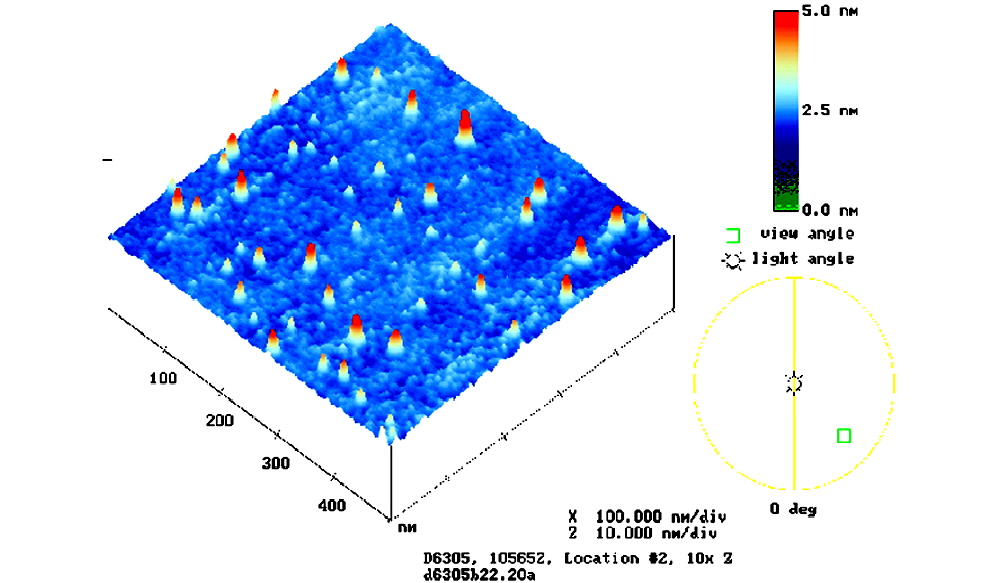
Figure 2. Atomic Force Microscopy (AFM) analysis. Shown are AFM scans of the SuperClean Microarray Substrate by Arrayit ArrayIt Division, Sunnyvale CA. Data are coded to a rainbow intensity scale, such that red data represents 5.0 nm or 50 angstroms. The substrates have an average smoothness of 2.0 nm or 20 angstroms, which corresponds to about 10 silicon dioxide bonds.

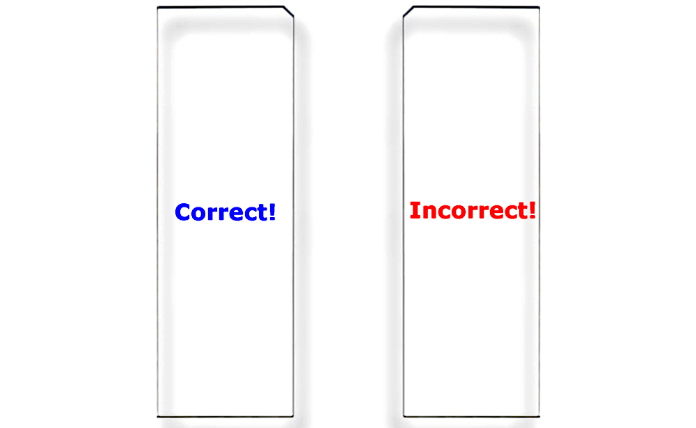
Figure 3. Correct Substrate Orientation. Shown is a graphic of two ArrayIt® Microarray Substrates, showing the correct and incorrect orientation for use. In the correct orientation (blue graphic), the chamfer will be located in the upper right corner and samples should be printed on the side facing upward, which is the same side that contains the word “Correct!”. In the incorrect orientation (red graphic), the chamfer will be located in the upper left corner, placing the backside facing upward, which is the side that contains the word “Incorrect!”. Only one side of ArrayIt® Microarray Substrates is suitable for printing. Please print on the correct side only.
Technical Assistance
Please contact us if you have any comments, suggestions, or if you need technical assistance. By electronic mail: arrayit@arrayit.com(under the subject heading please type "ArrayIt® technical assistance"). By telephone: (408) 744-1331, Monday–Friday PST 9:00am - 4:30pm. Please remember that we want to hear about your successes!
Scientific Publications
Click here and here for recent scientific publications using ArrayIt® brand microarray products from Arrayit International, Inc for protein A/G experimentation.
Recommended Equipment and Reagents
NanoPrint™ 2 Microarrayers
SpotBot® 4 Personal Microarrayers
InnoScan® Microarray Scanners
Microarray Hybridization Cassettes
High Throughput Wash Station
Microarray High-Speed Centrifuge
Protein Printing Buffer
BlockIt™ Blocking Buffer
Microarray Air Jet
Microarray Cleanroom Wipes
PCR Purification Kits
Micro-Total RNA Extraction Kit
MiniAmp mRNA Amplification Kit
Indirect Amino Allyl Fluorescent Labeling Kit
Universal Reference mRNA
Green540 and Red640 Reactive Fluorescent Dyes
Hybridization Buffers

