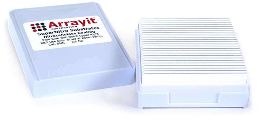
SuperNitro Nitrocellulose Membrane Coated
Data Sheet
![]() Shop this product in our online store
Shop this product in our online store
Products - Microarray Substrates & Slides - SuperNitro Nitrocellulose Membrane Coated Microarray Substrate Slides for DNA, Protein and RPPA Microarray Manufacturing

ArrayIt® SuperNitro Microarray Substrates offer highly innovative membrane surfaces for researchers wishing to utilize the time-tested attributes of nitrocellulose in a microarray format. SuperNitro can be used to attach DNA, protein, carbohydrate and any other molecule that has been shown to bind nitrocellulose in traditional filter assays. The high binding capacity of the 150 µm nitrocellulose layer is complimented by a polished atomically flat glass substrate for optimal performance compatibility with all standard glass substrate slide microarrayers and scanners.
NOTE: These substrates are compatible with AHC1x16, AHC1x24, AHC4x24, RC1x24 well reaction tools to multiplex the number of microarrays per silde substrate. Additionally the gaskets on these tools can be cut to customize the microarray printing and reaction area.
Product Description
- Glass substrate slides 25 x 76 x 1 mm
- Atomically flat polished glass substrate
- Corner chamfer for side orientation
- 150 µm nitrocellulose layer
- High binding capacity of 2 µg protein per square millimeter
- Reaction area 25 x 76 mm
- High-density bound streptavidin protein (Cat. SMNS)
- Compatible with both colorimetric and fluorescence applications
- Compatible with Arrayit’s complete microarray product and services line
SuperNitro is compatible with the SpotLight Microarray Scanner and the SpotWare Colorimetric scanner. The following detection methods can be used with this surface:
- Fluorescence
- Colorimetric
- Chemiluminescence
- Radioactivity
SuperNitro Microarray Substrates can also be partitioned into multiple wells of 2, 8, 12, 16, and 48 well formats for multiple microarrays on a single SuperNitro Substrate. These multiple well formats have all of the advantages of a single substrate with the exciting additional features:
- Increase the number of microarrays on each substrate
- Reduced cost per assay
- Individual wells dramatically reduce required sample volumes
The multiple wells on a single slide perfect for the following applications:
- Multiplexing experiments
- Side-by-side comparisons
- Control experiments
SuperNitro substrates are ready to use right out of the box without any additional processing or surface preparation. This surface is fully compatible with a variety of manufacturing technologies including ArrayIt® contact printing as well as non-contact printing such as ink-jetting. Protein Printing Buffer increases protein stability and provides extended printed microarray stability with increased signal compared to PBS buffers. BlockIt Blocking Buffer should be used for low background and to prevent non-specific binding in complex sandwich assays and other binding reactions. SuperNitro wash steps can be performed using 1X PBS buffers in a high throughput wash station.
SuperNitro Usage Tips
Printing the Microarray:
Proteins should be spotted in Protein Printing Buffer. If the protein is already in a 1X PBS solution, add an equal volume of printing buffer to achieve a final protein concentration between 0.5 and 2.0 mg/ml. SuperNitro is a 150-micron thick layer of pure nitrocellulose that has a much higher binding capacity than any other protein microarray surface available. When using a Robotic Microarrayer such as the NanoPrint, SpotBot 2 or other microarrayer in conjunction with an ArrayIt Spotting Device, decrease Z-axis speeds to 2 cm/sec to avoid damaging the nitrocellulose membrane. 45 to 65% humidity and ambient temperatures should be used. For best source sample integrity, use a system like the SpotBot or NanoPrint with source plate cooling.

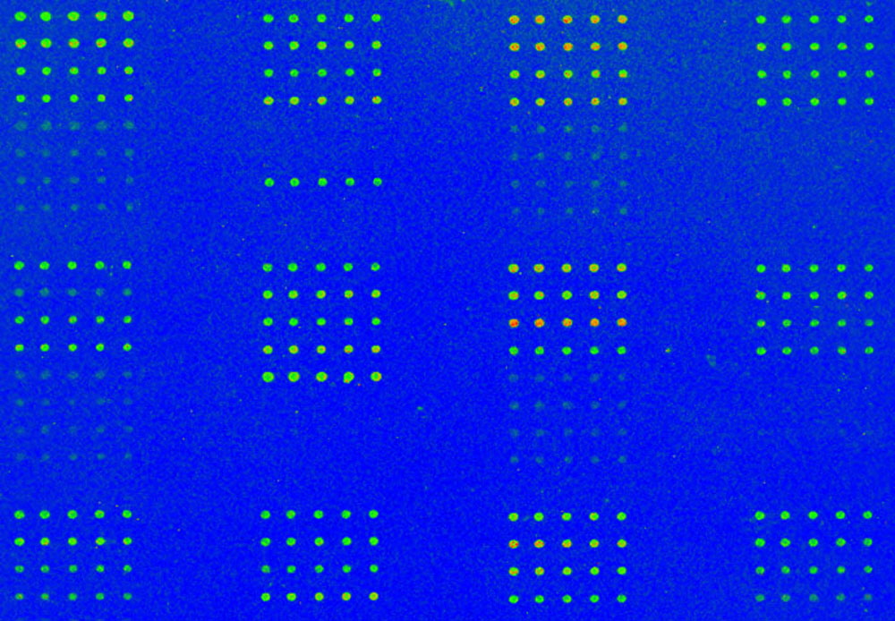
Figure 1. SuperNitro Microarray Substate Slides were printed with protein extract samples to form 150 µm diameter spots and stained with a fluorescent antibody for detection. Printing was performed using a NanoPrint™ Enterprise Level Protein Microarrayer with a professional series printhead and 946MP4 microarrray printing pins. Scanning utilized an InnoScan® 710AL Two-Color Flourescence Microarray Scanner at 5 µm scan resolution and gain 10. Data analysis was acheived using MAPIX® Quantification Software.
Mask configurations are available in 1, 2, 4, 8, 12, 16 and 48-well sizes, allowing multiple samples to be tested on a single SuperNitro Substrate. Hydrophobic masks greatly reduce the cost of SuperNitro usage, which is particularly helpful for patient testing and diagnostic applications. A Microarray Imprinter can be used on this surface.
Printed Microarray Storage:
Printed microarrays should be incubated overnight at low humidity to establish the coupling reaction. Arrays can be stored in a clean, dry cool place. Refrigeration can be used, but Protein Printing Buffer is designed to stabilize proteins for long-term storage at room temperature. Vacuum packing can also be used; make sure microarrays are dry prior to vacuum packing. Store printed microarrays unblocked and un-washed, performing blocking and washing steps just prior to incubation.
SuperNitro Blocking & Washing:
Prior to performing binding reactions, SuperNitro should be blocked with BlockIt Buffer for 1 hour and then washed 3 times in 1X PBS for 5 minutes using a high throughput wash station. Do not let the surface dry out between blocking, binding and washing reactions. SuperNitro should only be allowed to dry after printing and prior to scanning only. If a multiple well slide is being used, when washing, be sure to flood the microarray with sufficient volume of 1XPBS to avoid cross contamination. Use of an ArrayIt high throughput wash station will ensure success.
SuperNitro Incubation and Detection Reactions:
Binding reactions can be 1 hour to overnight. Experiments must not be allowed to dry
out. Fluorescent reactions running for long periods of time should be kept in the dark. Secondary antibody reactions should be run for up to 2 hours and typically not less than 30 minutes. Concentration of detection antibody needs to be optimized empirically by the user. Colorimetric and other enzymatic detection reactions require optimization by the user.
Scanning:
SuperNitro substrates are compatible with a variety of detection methods. The microarray should not be detected wet or damp unless the detection method is chemiluminescent. Air-dry the slide in a dark, dust free place until the membrane appears white. When imaging SuperNitro and other white surfaces in laser/pmt microarray scanners, the default laser/PMT for glass transparent surfaces are not optimal for best signal to noise ratio. Because of the increased binding capacity of the nitrocellulose 3-D layer lower laser power and PMT settings should be used. For best signal to noise rations in fluorescent detection use the SpotLight Microarray Scanner.

Figure 2. Shown are background fluorescence scans of Arrayit SuperNitro Microarray Substrate Slides (left) and another leading brand (right). Note the SuperNitro has 5 times lower intrinsic background compared to the other leading brand. Low intrinsic fluorescence is essential for high detectivity and sensitivity in microarray assays.
Scientific Publications
Click here for recent scientific publications using Arrayit SuperNitro Microarray Substrate Slides.
Recommended Equipment and Reagents
NanoPrint 2 Microarrayers
SpotBot® 4 Personal Microarrayers
SpotBot® Extreme Microarray Spotters
SpotBot® Titan Microarray Arrayers
InnoScan® Microarray Scanners
SpotLight 2 Microarray Scanners
Microarray Hybridization Cassettes
High Throughput Wash Station
Microarray High-Speed Centrifuge
Protein Printing Buffer
BlockIt Blocking Buffer
Microarray Air Jet
Microarray Cleanroom Wipes
PCR Purification Kits
BlockIt Blocking Buffer
Micro-Total RNA Extraction Kit
MiniAmp mRNA Amplification Kit
Indirect Amino Allyl Fluorescent Labeling Kit
Universal Reference mRNA
Green540 and Red640 Reactive Fluorescent Dyes
Hybridization Buffers

