Hydrogel Coated Three-Dimensional Microarray Slides
Data Sheet
![]() Shop this product in our online store
Shop this product in our online store
Products - Microarray Substrates & Slides - Hydrogel Coated Three-Dimensional Microarray Slides

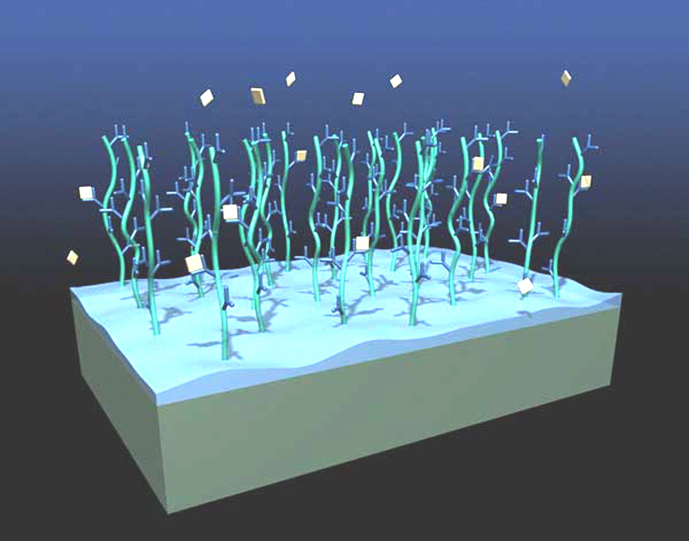
ArrayIt® and XanTek have teamed up to provide a series of premium hydrogel glass substrate slides for microarray applications. Based on nearly 10 years experience in surface manufacturing biosensors, XanTec bioanalytics has designed a novel hydrogel coating with drastically enhanced protein immobilization capacity without showing the typical disadvantages of thick film coatings. Compared to many surface coatings, the immobilization capacity of HC-series hydrogel matrix is 10 – 50 times higher; typical protein binding capacity is in the range between 10 - 100 ng/mm². The polymer chains on these hydrogel coatings are oriented in a brush-like manner perpendicular to the surface allowing rapid diffusion through the 5 µm thick layer. Hydrogel microarray surfaces support.
Product Description
The biocompatible character of the HC hydrogel slides is similar to separation media used in protein purification and stabilizes even sensitive proteins, thus ensuring excellent long term storage stability. Another interesting feature is the screening of fluorophor markers in the dry state. Bleaching, dimerization and quenching phenomena are suppressed and the fluorophor signal is maximized.
A straightforward, robust spotting protocol and the efficient immobilization process yields consistent high S/N ratios already at low protein quantities, saving valuable biomaterial. In a typical spotting experiment, approximately 50% of the spotted protein is immobilized with a typical CV of < 10% over the slide surface. The contact angle of the activated surface is 50° - 55° which is a good prerequisite for homogeneous spot shapes. After quenching, the surface is very hydrophilic (θ=0°). Blocking steps are usually not required, significantly enhancing the biospecificity of the resulting protein microarrays.
Due to the covalent immobilization, small ligands such as peptides, cytokines and other low MW compounds can also be immobilized in a stable manner, provided that they bear amino or other functional groups for covalent coupling.

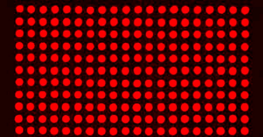
Figure 2. Uniform spot morphology of printed Oyster® 656 fluorescently labeled BSA on a hydrogel surface.

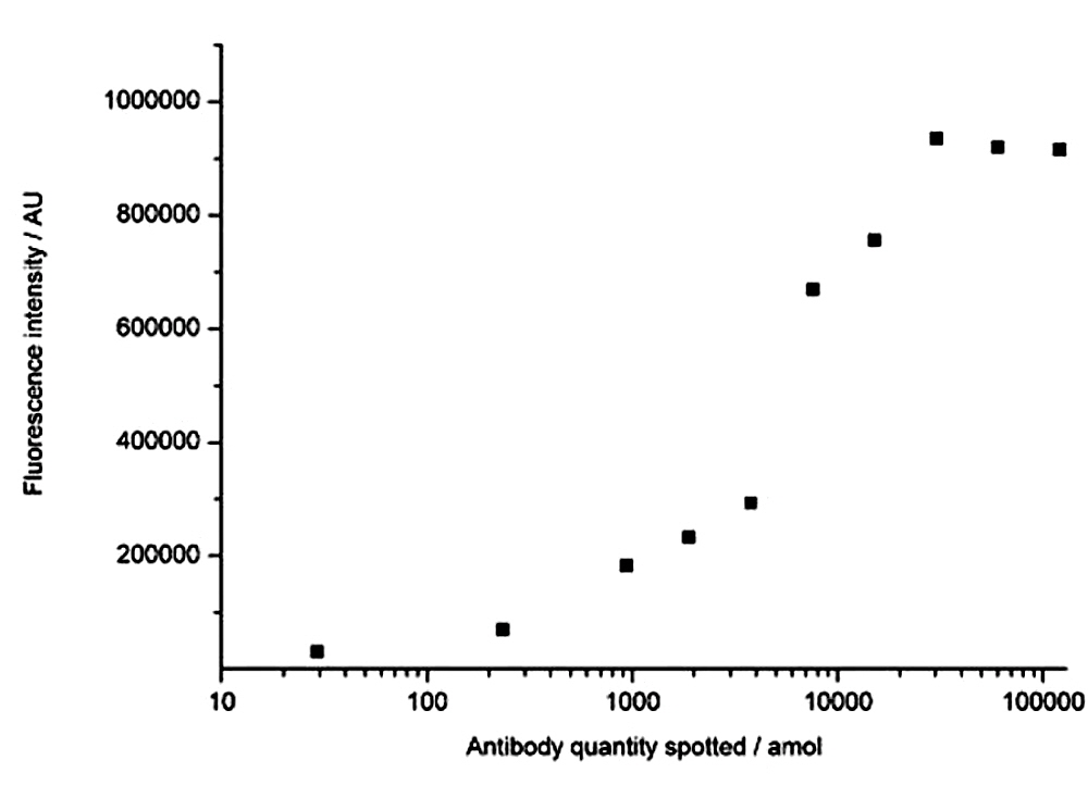
Figure 3. Evalutation of immobilization efficiency. Different quantities ofC-reactive protein antibody were spotted onto NHS-activated carboxymethyldextran (similar to the HC surface) slides followed by quenching and overnight incubation with 10 µg Cy5-CRP per ml in PBS. Scanning was performed with a GMS 418 scanner. Spots above the upper detection limit at 65.000 AU were scanned at lower gains and recalculated accordingly. The average background noise was approximately 1,500. The lowest concentration of 27 amol per spot yielded a signal of 31,000 and thus a signal to background ratio of 20. At 30 fmol per spot the maximum immobilisation capacity was observed with a corresponding signal of 850,000 and a signal to background ratio of 570.
Scientific Publications
Click here and here for recent scientific publications using ArrayIt® brand microarray products from Arrayit International, Inc for hydrogel experimentation.
Recommended Equipment and Reagents
NanoPrint™ 2 Microarrayers
SpotBot® 4 Personal Microarrayers
InnoScan® Microarray Scanners
SpotLight™ 2 Microarray Scanners
Microarray Hybridization Cassettes
High Throughput Wash Station
Microarray High-Speed Centrifuge
Protein Printing Buffer
BlockIt™ Blocking Buffer
Microarray Air Jet
Microarray Cleanroom Wipes
PCR Purification Kits
SuperProtein Blocking Buffer
Micro-Total RNA Extraction Kit
MiniAmp mRNA Amplification Kit
Indirect Amino Allyl Fluorescent Labeling Kit
Universal Reference mRNA
Green540 and Red640 Reactive Fluorescent Dyes
Hybridization Buffers

