Protein Microarray Buffers
Data Sheet
![]() Shop this product in our online store
Shop this product in our online store
Arrayit | PMBK PPB activation blocking wash rinse printing buffer solutions DNA protein microarray life sciences research
Reagents - Microarray Buffers - Protein Microarray Buffers - Protein Printing Buffer Plus to Enhance Protein Microarray Control Sample Manufacturing

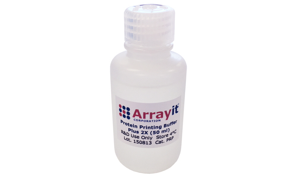
Arrayit advanced formulation for peptide and protein microarray manufacturing that enhances the precision and printing quality of protein dilution series controls such as IgG, anti-IgG and other sample types essential for microarray testing.
Use Protein Printing Buffer Plus on SuperEpoxy 2 and 3 glass substrate slide surfaces when printing dilution series of control proteins. This buffer is delivered at a 50 ml of 2X solution.
Table of Contents
- Introduction
- Quality Control
- Product Description
- Technical Note
- Technical Assistance
- Short Protocol
- Complete Protocol
- Recommended Equipment
- Troubleshooting Tips
- Ordering Information
- Warranty
Introduction
Congratulations on taking a big step towards improving the economies of scale, quality and speed of your proteomics research. This booklet contains a complete set of protocols outlining the steps and principles needed to use Arrayit Protein Printing Buffer.
Product Description
The Arrayit Protein Printing Buffer Plus (PRP) is an advanced buffering system containing a proprietary mixture of ionic and polymeric materials. A specially formulated buffer for microarray manufacturing of dilution series control proteins on SuperEpoxy 2 and 3 Microarray Surface Chemistry. Not for use on membrane surfaces.
Users will appreciate the following features of Protein Printing Buffer Plus (PRP):
- Supports Arrayit’s most widely used printing technology
- Print proteins, enzymes, antibodies, receptors, antigens, and peptides
- Washes away during blocking and processing
- Manufacture microarrays of pure proteins, recombinant proteins and cellular extracts
- Stabilizes protein samples and prevents denaturation
- Provides uniform feature size and coupling density accross lowering concentrations
- Protects printed samples from environmental damage
- Slows sample evaporation within the source microplates
- Minimizes sample drying and crystallization on substrates and pins
- Washes away easily, leaving pure bound protein molecules
- Improves deposition uniformity which facilitates data analysis
- Arrives pre-mixed as a 2X solution, sterile and protease-free
- Sufficient to print 50 million protein features (5,000 10K protein arrays)

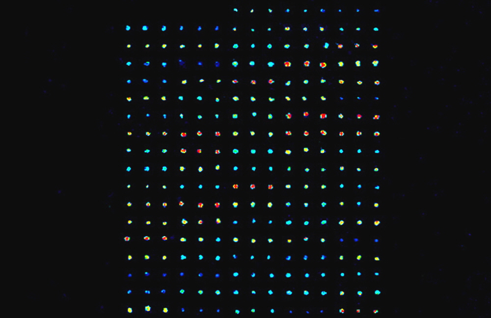
Figure 2. Ten Fold Dilution series of Human IgG starting at 1 µg/µl Printed in Protein Printing Buffer Plus left to right top down. Microarrays manufactured using a NanoPrint Microarrayer, Professional Printhead and 946MP3 Microarray Printing Pins. SuperEpoxy2 Microarray Substrates, Blocked wtih BlockIt Plus, reacted with Anti-Human IgG Conjugated to Cy3 using diluted 1 to 2000 in Protein Microarray Reaction Buffer using an RC1x24 Microarray Reaction Cassette. Microarray Scan at 5 um resolution using an Innoscan 710 Microarray Scanner.
Technical Note
It may be necessary to add protease or phosphatase inhibitors to the Protein Printing Buffer to increase the stability of certain proteins or protein extracts. Protein stability can also be enhanced if microarray manufacture is performed at 4°C. The SpotBot and NanoPrint Microarrayers can be operated in a cold room. A special “cold platen” is also available for these machines if a 4°C printing temperature is required in an ambient laboratory environment. Please contact arrayit@arrayit.com for technical details.
Technical Assistance
Please contact us if you have any comments, suggestions, or if you need technical assistance. By electronic mail: arrayit@arrayit.com (under the subject heading, please type, “Technical assistance”).
Short Protocol (Steps 1-10)
1. Obtain 0.2-1.0 µg/µl protein samples.
2. Transfer 4.0 µl per well of each protein sample into 96- or 384-well microplates.
3. Add 4.0 µl per well of 2X Arrayit Protein Printing Buffer (PPP).
4. Mix the samples by pipetting up and down 10 times.
5. Print protein samples onto SuperEpoxy 2 or 3 .
6. Use BlockIt Plus to block and process the printed protein microarrays.
7. React the processed microarrays with fluorescent samples.
8. Wash the microarrays to remove unreacted fluorescent material.
9. Scan the microarray to produce a fluorescent image.
10. Quantitate and model the fluorescent data.
Complete Protocol (Steps 1-10)
1. Obtain 0.2-1.0 µg/µl protein samples and determine the desired dilution series. Phosphate buffered saline (PBS) at 1X concentration works very well for most proteins. Protein samples should be free of aggregates and particulates that can clog printing devices and impair attachment to the microarray substrate. Aggregates and particulates can be removed by centrifugation or filtration. A 50kD protein at 1 µg/µl concentration has a concentration of 20 µM. At 30% coupling efficiency, a 20 µM protein will produce a target density of 1011 proteins per mm2 of substrate.
2. Transfer 4.0 µl per well of each protein sample into 96- or 384-well microplates. Transfer can be performed manually by pipette or with a liquid handling robot. Certains proteins are fragile and protein samples should be handled with care to avoid damaging the structure and function of proteins.
3. Add 4.0 µl per well of 2X Arrayit Protein Printing Buffer (PRP). This will give a final concentration of 1X Protein Printing Buffer and a final protein concentration of 0.1-0.5 µg/µl. Certain proteins or protein extracts are more stable at 4°C. Keeping the proteins samples cool may improve stability. Stability can also be improved in some cases by the addition or protease and phosphatase inhibitors or by the use of a SpotBot or NanoPrint Microarrayer equipped with a cooled platen.
4. Mix the samples by pipetting up and down 10 times. This step ensures that the protein samples mix thoroughly with the 2X Protein Printing Buffer Plus. Failure to mix the samples thoroughly will produce poor quality microarrays.
5. Print dilution series onto SuperEpoxy 2 or 3 Microarray Substrates. The SuperEpoxy surface couples proteins more readily than other surfaces owing to the increased reactivity of the epoxide groups. Another advantage of SuperEpoxy is that coupling can occur in humid and conditions as may be required to maintain protein structure and function.
6. Use BlockIt Plus blocking buffer and process the printed protein microarrays. Once the printing process is complete, wash the printed microarrays Protein Microarray Wash Buffer to remove unbound protein molecules and components of the printing buffer. Protein binding to the SuperEpoxy 2 and 3 surface is extremely stable and the microarrays can be washed, blocked and reacted without sufficient loss of coupled protein. Allow microarrays to dry overnight to maximize binding. A good blocking protocol involves a 1 hour room temperature to overnight incubation at 4 degrees C in 1X BlockIt Plus, using a High Throughput Wash Station or petri dish. Blocking can be performed with or without agitation. The BlockIt Plus blocking step will inactivate unreacted epoxy groups and prevent background noise. After blocking, wash the microarrays in Protein Microarray Wash Buffer follow by a short few second rinse in Protein Microarray Rinse Buffer. Use High Throughput Wash Station, petri dish or similar device to get gentle agitation for good washing.
7. React the processed microarrays with fluorescent samples. Processed microarrays containing coupled target proteins can be reacted with fluorescent samples to study protein-protein interactions. Binding reactions can be performed using Protein Microarray Reaction Buffer. Fluorescent samples can be incubated as a droplet over the printed microarray, underneath a cover slip, or in a microfluidics chamber. Consider using AHC A 60-minute incubation at room temperature is usually sufficient to obtain strong binding and intense fluorescent signals (see Fig. 1). A Hybridization Cassette can be used to prevent sample evaporation during prolonged binding reactions.
8. Wash the microarrays to remove unreacted fluorescent material. Once binding between the bound target proteins and the fluorescent protein probe molecules is complete, wash the microarray to remove the unbound material. Washes can be performed three times for 5 min each at room temperature in 1X PBS. After the wash procedure, excess buffer should be removed from the surface by tapping or by centrifugation with a Microarray High-Speed Centrifuge.
9. Scan the microarray to produce a fluorescent image. The fluorescent microarray can be scanned or imaging using any of a number of high quality commercial detection instruments, but for best results use Innoscan Microarray Scanners. Instrument settings can be adjusted to optimize the imagine acquistion process.
10. Quantitate and model the fluorescent data using Mapix or other microarray quantification software.
Recommended Equipment
NanoPrint 2 Microarrayers
946 Spotting Device
Stealth Micro Spotting Device
SpotBot® 4 Personal Microarrayers
SuperEpoxy 3 Microarray Substrates
Protein Printing Buffer
BlockIt Blocking Buffer
High Throughput Wash Station
Microarray High-Speed Centrifuge
Microarray Hybridization Cassette
Troubleshooting Tips
Poor printing quality:
- Incomplete mixing of protein samples in Protein Printing Buffer Plus
- Poor printing environment (50% humidity and 25°C recommended).
- Check wash dry station of microarrayer to make sure it is functioning
- Damaged microarary pritning pins
- Not using SuperEpoxy 2 or 3 Surface Chemistry
Poor protein coupling:
- Poor surface chemistry (Use SuperEpoxy 2 or 3)
- Inhibitor in protein sample (suspend in 1X PBS and then 1X PRP)
Weak fluorescent signals:
- Fluorescent protein probe mixture binds poorly to protein targets
- Probe labeling inefficient
- Washes too harsh
Reagents - Microarray Buffers - Protein Microarray Buffers to Enhance Protein Profiling, Peptide, Serum IgG and IgE, and Auto-Antibody Protein Microarray Assays

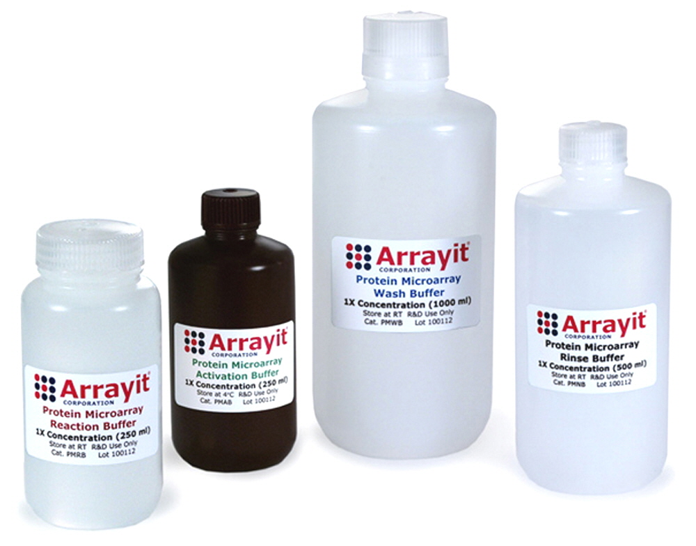
Arrayit’s Protein Microarray Kit is the first complete protein microarray buffer system on the market. Kit contents include activation buffer, reaction buffer, wash buffer and rinse buffer. Supports all protein microarrays and takes the guess work out of microarray-based proteomic studies. Buffers are 0.1 µm-filtered, pre-mixed and ready to use. Buffers increase signal strength and reduce background. Highly recommended for peptide, antibody, antigen, reverse phase and PlasmaScan™ Microarrays. We also offer reaction and blocking buffer automation formulations for high-throughput research and screening applications.
Table of Contents
- Introduction
- Quality Control
- Product Description
- Kit Contents
- Technical Assistance
- Short Protocol
- Complete Protocol
- Troubleshooting Tips
- Recommended Products
- Ordering Information
- Warranty
Introduction
Arrayit Protein Microarray Buffers improve the precision, speed and affordability of your microarray research in proteomics, pharmaceuticals, diagnostics, agriculture, and other areas of life sciences and health care research. This handbook contains all the information required to take full advantage of Arrayit Protein Microarray Buffers.
Quality Control
Arrayit uses the highest quality control (QC) and quality assurance (QA) measures to ensure the quality of our Arrayit brand Protein Microarray Buffers. The finest scientific research was used to develop this product line. The use of advanced R&D, ultra-pure reagents, and 0.1 µm filtration guarantees that this product line outperforms the highest industry standards.
Product Description
Arrayit Protein Microarray Buffers are designed for all protein microarray applications. Users will appreciate the following features:
- Based on the finest microarray research and development (R&D)
- Excellent for protein expression and serum diagnostics applications
- Designed for proteins, antigens, peptides, cell extracts and serum samples
- Promote strong signal intensities and high binding specificity
- Superior formulations reduce background
- Each lot checked by quality control (QC) and quality assurance (QA)
- Ultra-pure reagents used for buffer formulation
- All buffers purified by 0.1 µm filtration
- Provided in convenient bottles as 1X concentrates
- Each kit will process up to 100 protein microarrays
- Arrive pre-mixed and ready to use
- Compatible with protein microarrays from Arrayit and other vendors
- Remove the guesswork from protein microarray applications
Kit Contents
The Arrayit Protein Microarray Buffer Kit contains the following components:
- 250 ml of 1X Protein Microarray Activation Buffer
- 250 ml of 1X Protein Microarray Reaction Buffer
- 1000 ml of 1X Protein Microarray Wash Buffer
- 500 ml of 1X Protein Microarray Rinse Buffer
Technical Assistance
Please contact us if you have any questions, comments, suggestions, or if you need technical assistance. By electronic mail, use arrayit@arrayit.com and type "ArrayIt technical assistance" into the subject line. By email, arrayit@arrayit.com between the hours of 8AM and 7PM PST Monday through Friday. We want to hear about your successes and are always happy to feature contributed data on our website.
Complete Protocol (Steps 1-5)
1. Activate Microarrays. Protein microarrays require activation prior to use to remove unbound protein molecules from the surface and to shield the surface against non-specific binding. Activate protein microarrays by mixing for 60 min in Protein Microarray Activation Buffer (Cat. PMAB). The activation step can be performed with gentle mixing in 15 ml of Protein Microarray Activation Buffer in a petri dish or 450 ml of Protein Microarray Activation Buffer in a High-Throughput Wash Station. Following activation, wash the microarrays two times for 5 min in Protein Microarray Wash Buffer (Cat. PMWB) and one time for 1 sec in Protein Microarray Rinse Buffer (Cat. PMRB). All washes can be performed using 25 ml volumes in a petri dish or 450 ml volumes in a High-Throughput Wash Station (Cat. HTW) with gentle agitation. After washing, spin for 1 sec in a Microarray High-Speed Centrifuge to mostly dry the microarrays and proceed immedately to the next step.
2. React the activated microarrays with samples. Samples can be are prepared by mixing 5-25 µg of protein with Protein Microarray Reaction Buffer (Cat. PMRB), making sure not to dilute the Protein Microarray Reaction Buffer by more than 1.5-fold. The same volume of protein sample should be used for all reactions in which comparative information is sought. Reactions can be performed using LifterSlip™ (elevated cover slips) in a 15-75 µl volume (depending on the size of the LifterSlip™) or multi-well reaction cassettes Catalog ID’s AHC1x16, AHC1x24 and AHC4x24 for using an 80 µl per-well volume and 50 µl for the RC1x24 reaction cassette. Binding reactions may also be performed using large sample droplets or a variety of custom gaskets. Reaction areas can be made using the Microarray Imprinter. For example, undiluted or diluted biotinylated protein samples can be used depending on the sample concentration and plasma protein expression level. Typically 5 µg of labeled protein is sufficient to obtain strong signals. Up to 25 µg of labeled protein can be used, though saturating signals and non-linearity can be observed at high sample protein concentrations. Fluorescent samples can be reacted in the same manner as biotinylated samples. Typical binding reactions run for 60 to 90 min at room temperature with gentle mixing. Following the reaction step, wash the microarrays three times for 3 min in Protein Microarray Wash Buffer (Cat. PMWB) with gentle agitation. Washes can be performed using 25 ml volumes in a petri dish or 450 ml volumes in a High-Throughput Wash Station with gentle agitation. After washing, spin for 1 sec in a Microarray High-Speed Centrifuge to dry the microarrays.
3. Stain the microarrays with a fluorescent secondary reagent. Prepare the secondary antibody reagent by diluting the fluorescent conjugate to a final concentration of 1 µg/ml in Protein Microarray Reaction Buffer (Cat. PMRB). For example, a one thousand-fold (1:1,000) dilution of the 1 mg/ml Cy3™-Streptavidin staining reagent from Jackson ImmunoResearch (Cat. 109-165-088) works well. Other secondary antibodies can be used to measure IgG, IgE, IgM and IgA. Antibody pairs can be used in sandwich assays. A 1.0 ml volume of secondary reagent is prepared by mixing 1.0 µl of secondary antibody in 1 ml of Protein Microarray Reaction Buffer. Mix by inverting the tube 10 times. Stain the microarrays using 25-125 µl of diluted secondary reagent. The staining volume will depends on whether a single or multi-well microarray format is being used. Stain for 60 min at room temperature with gentle agitation. Following the staining step, wash the Pmicroarrays three times for 1 min in Protein Microarray Wash Buffer (Cat. PMWB) and one time for 1 sec in Protein Microarray Rinse Buffer (Cat. PMNB) with gentle agitation. Washes can be performed using 25 ml volumes in a petri dish or 450 ml volumes in a High-Throughput Wash Station with gentle agitation. After washing, spin for 1 sec in a Microarray High-Speed Centrifuge to dry.
4. Scan the microarrays for fluorescence emission. Scan the PlasmaScan™ Antibody Microarrays using an Arrayit SpotLight™ (Cat. SLMS) or Arrayit InnoScan® 710 (Cat. 710) or 900 (Cat. 900) microarray scanner set to detect Cy3™ or Cy5™. PlasmaScan™ can also be read with other brands of microarray scanner compatible with the open platform substrate slide 25 x 76 mm format. Scanner settings should be adjusted to minimize saturated signals to 1% or less. Multiple scans can be taken to capture data at different sensitivity levels. All data files should be saved as 16-bit TIFF images for data analysis.
5. Quantify and model the fluorescent data. Launch Arrayit Mapix Microarray Quantification Software and open the Microarray TIFF. Create a quantification grid by importing the GAL file provided by the NanoPrint Microarrayer. Adjust the grid so that it fits approximately around each printed element. Once the grid is aligned, click “find spots automatically” to allow automated spot finding by Mapix. Minor adjustments to individual spot locations can be made using the “spots modification” command. Click on “quantification process” to quantify the PlasmaScan™ spot intensities. Click on the “data viewer” in the “Window” menu to visualize the quantified data. Click on “Save results” to save the data as a text file (.txt). The .txt file can be imported into spread sheets and other software programs for additional analysis.

Figure 1. Protein microarray data obtained using the Arrayit Protein Microarray Buffer Kit (Cat. PMBK). Monoclonal antibodies were printed in triplicate as 75 µm features onto SuperEpoxy 2 Microarray Substates (Cat. SME2), activated with Protein Microarray Activation Buffer, reacted with Protein Microarray Reaction Buffer containing biotin-labeled human serum, stained with Cy3-labeled streptavidin, washed with Protein Microarray Wash Buffer and rinsed with Protein Microarray Rinse Buffer prior to scanning. The fluorescent image indicates strong signal intensities and low background.
Troubleshooting Tips
High background
- Increase stringecy of wash with agitation prior to binding reaction
- Increase stringency of blocking by elevating temperature to 37C
- Fluorescent detection reagents are drying out during detection step.
- Keep dry steps short in between reactions, run reactions all the way through without stopping once the protocol is started.
Scientific Publications
Click on the links for recent scientific publications using Arrayit brand products for protein microarray experimentation.
Recommended Products
- Hybridization Cassettes
- High-Throughput Wash Station
- Microarray High-Speed Microarray Centrifuge
- Arrayit Blocking Buffers
- Nanoprint™ Microarrayers
- 946 Microarray Printing Technology
- SpotBot® 3 Personal Microarrayers
- PCR Purification Kits
- InnoScan® Microarray Laser Scanners
- SpotLight™ 2 Microarray Scanners
- SpotWare™ Colorimetric Microarray Scanners

