Alkaline Phosphatase Labeling Kits
Data Sheet
![]() Shop this product in our online store
Shop this product in our online store
Arrayit | Alkaline phosphatase kits APK colorometric microarrays life sciences research
Reagents - Microarray Labeling - Alkaline Phosphatase Kits for DNA and Protein Microarrays in Research, Life Sciences, Molecular and Pharmaceuticals
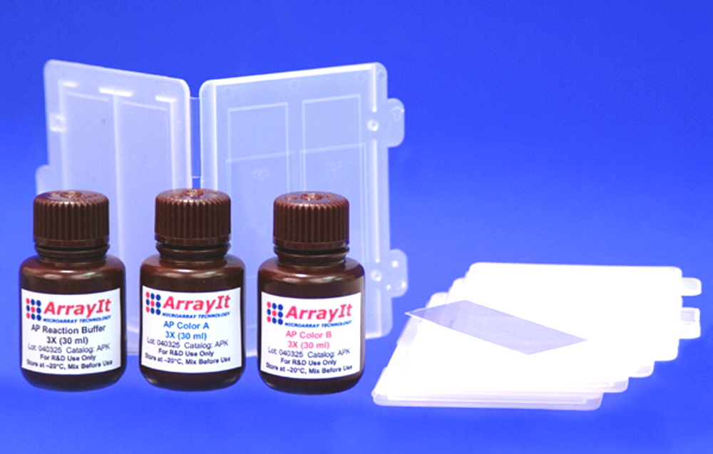
Arrayit Alkaline Phosphatase Kit opens up entirely new avenues of research and diagnostics for DNA and protein microarrays and proteomics. ArrayIt® sets the standard for innovative colorimetric microarray solutions.
Table of Contents
- Introduction
- Quality Control
- Product Description
- Kit Contents
- Short Protocol
- Complete Protocol
- Technical Assistance
- Troubleshooting Tips
- Recommended Products
- Ordering Information
- Warranty
Introduction
Congratulations on taking a big step towards improving your protein microarray research. This booklet contains complete protocols outlining the steps required to use the ArrayIt® Alkaline Phosphatase Kit.
Quality Control
TeleChem assures the performance of this ArrayIt® product. The highest levels of scientific research and innovation were used to develop this kit.
Product Description
ArrayIt® provides the world’s only Alkaline Phosphatase Kit for microarray research. This innovative hardware and assay solution makes alkaline phosphatase (AP) assays rapid, affordable and easy to implement. Users will appreciate the following features:
- Contains AP developing reagents and assay hardware
- Novel blue colorimetric chemistry increases reagent and pigment stability
- Light-proof bottles prevent photo-degradation
- Develops any alkaline phosphatase microarray
- Use on membrane-coated and silane-based glass surfaces
- Rapid reaction time of 1-30 min
- Total assay time of 60 min
- >1000-fold dynamic range
- Extremely high assay sensitivity and low background
- Highly affordable at $0.27 per microarray
- Hardware can also be used for upstream protein and antibody binding steps
- Convert any conventional AP assay into a microarray format
- Environmentally friendly formulation contains no organic solvents
- 100 reactions per kit
Kit Contents
The ArrayIt® Alkaline Phosphatase Kit (APK) contains the following components:
- Five (5) dual substrate reaction cassettes
- Five (5) 24 x 60 mm glass cover slips
- One (1) 30 ml bottle of 3X AP Reaction Buffer
- One (1) 30 ml bottle of 3X AP Color A
- One (1) 30 ml bottle of 3X AP Color B
Short Protocol (steps 1-7)
1. Place alkaline phosphatase microarray into reaction cassette.
2. Prepare 300 µl of AP Developer.
3. Transfer 125 µl AP Developer onto the microarray.
4. Add cover slip and incubate 1-30 min at RT.
5. Stop the reaction by flooding cassette with dH2O.
6. Spin dry by centrifugation.
7. Scan microarray to detect blue colorimetric signals.
Complete Protocol (steps 1-7)
1. Place alkaline phosphatase microarray into reaction cassette. Prepare any microarray bound with an alkaline phosphatase reagent such as antibody-AP conjugate. Place the two microarrays “spots up” in the bottom portion of the plastic reaction cassette (Fig. 1). Make sure the substrates are seated firmly against cassette.
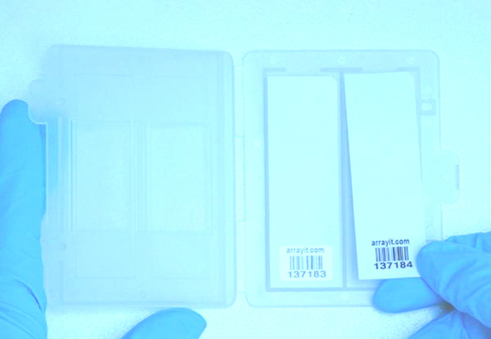
Figure 1. Placing two microarrays in the alkaline phosphatase reaction cassette.
2. Prepare 300 µl of AP Developer. Thaw all three bottles of reagents, which should be stored frozen at –20°C before and after use. After thawing, mix the reagents thoroughly before use by inverting and swirling the bottles 5-10 times with the caps tightly sealed. Failure the mix the reagents at this step will reduce assay performance. To a microfuge tube, add 100 µl 3X AP Reaction Buffer, 100 µl 3X AP Color A, and 100 µl 3X AP Color B, and mix thoroughly. This 300-µl mixture of AP Developer is sufficient to develop 2 microarrays (24 x 60 mm) at 125 µl per microarray.
3. Transfer 125 µl AP Developer onto the microarray. Using a 200-µl pipette, remove 125 µl of AP Developer from the 300 µl mixture and place the 125-µl drop directly onto the microarray (Fig. 2). Move quickly to Step 4 to avoid uneven AP developing.
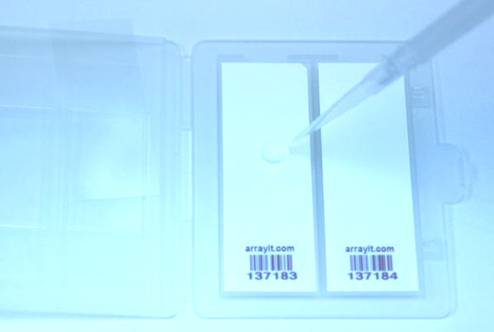
Figure 2. Pipetting 125 µl of AP developer onto the microarray surface. Note that for proper orientation the corner chamfer should be located in the upper right corner of the substrate. Move quickly to place the glass cover slip (left) onto the droplet (see Fig. 3) to avoid uneven developing.
4. Add cover slip and incubate 1-30 min at RT. After pipetting the 125-µl drop of AP Developer, immediately add a 24 x 60 mm cover slip directly onto the droplet, using a forceps to lower it slowly (Fig. 3). Care should be taken not to introduce air bubbles at this step. The AP Developer should sheet evenly across the entire area of the cover slip, forming a uniform 87-µm layer of reagent across the microarray. This reagent thickness provides a sufficient reagent concentration to develop any type of alkaline phosphatase microarray. The reaction can be incubated at room temperature (20-25°C) or at 37°C for accelerated reaction kinetics. The total incubation time is 1-30 min depending on the amount of AP enzyme on each microarray. One approach is to incubate one microarray for 1-5 min and a second microarray for 20-30 min, to obtain different “exposures” spanning a wide range of signal intensities corresponding to weak and strong signals. Some trial and error may be required to assess the correct development time. Excessive development time will lead to a loss of quantitation and elevated background, and should be avoided.
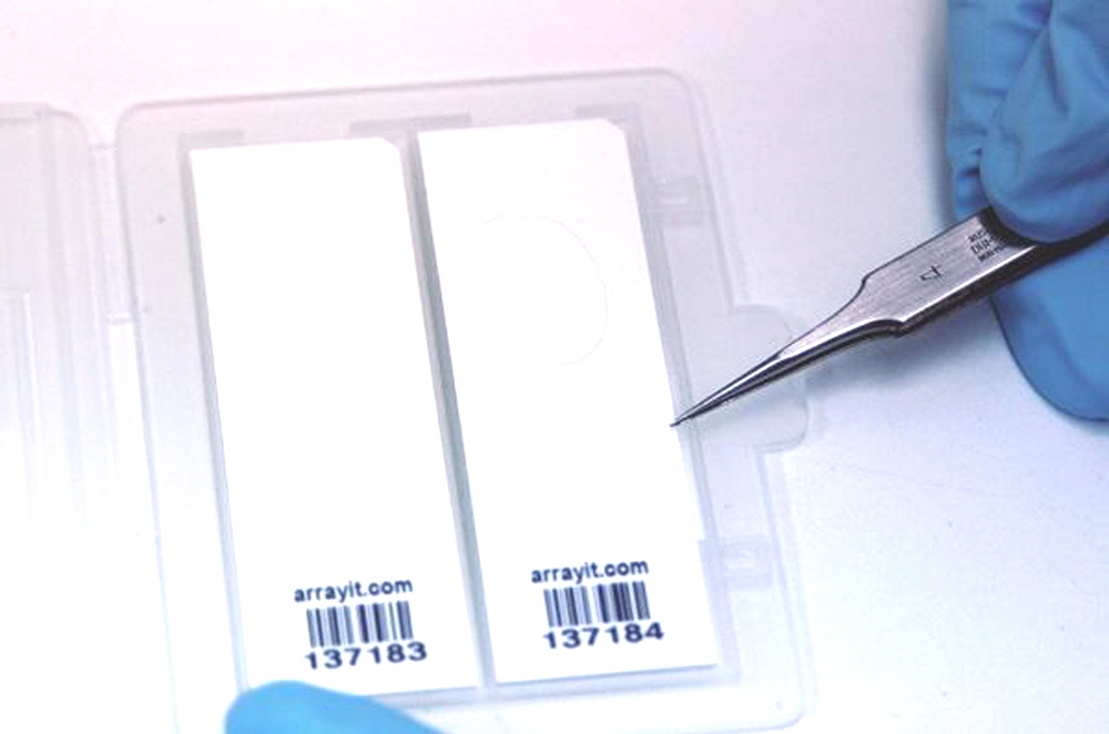
Figure 3. Placing a 24 x 60 mm glass cover slip onto a 125-µl droplet of AP Developing reagent.
5. Stop the reaction by flooding cassette with dH2O. After 1-30 min of developing time, carefully open the reaction cassette, secure the microarrays in position by applying light pressure between thumb and forefingers, and flood the surface of the microarrays with a steady stream of distilled H2O (Fig. 4). The glass cover slip should float free and fall off of the microarray at this step. Make sure the cover slip dislodges within the first 10 seconds of washing to prevent uneven developing. The reaction will be stopped completely after 30 seconds of washing. Close the cassette and flip it downward several times to remove residual dH2O (Fig. 5) to provide nearly dry microarrays that are ready for complete drying by centrifugation.
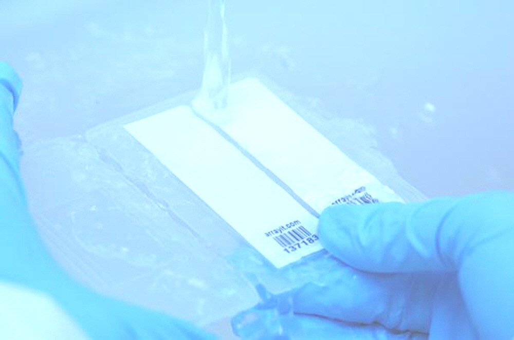
Figure 4. Stopping the alkaline phosphatase development reaction using a steady stream of distilled water.
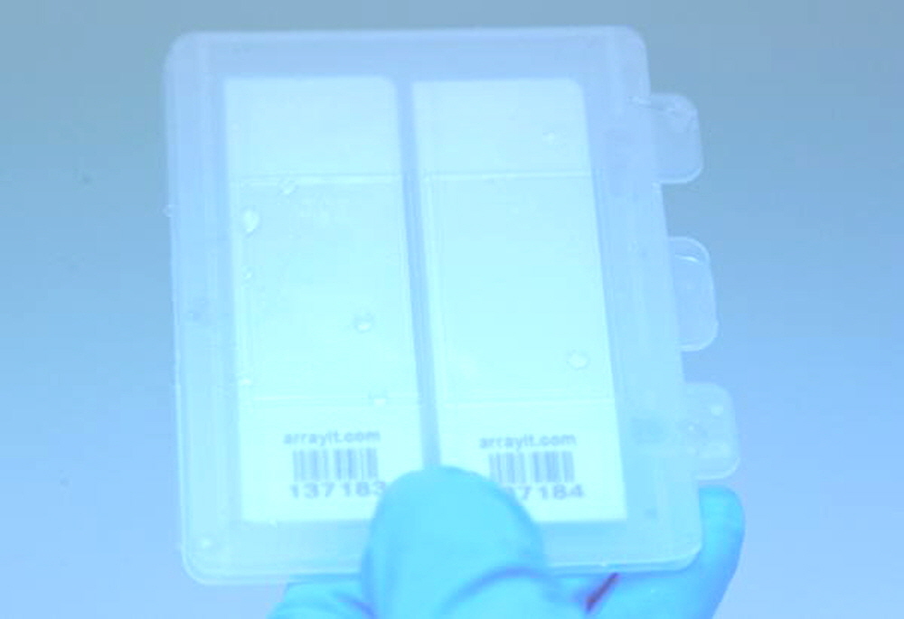
Figure 5. Removing residual distilled water after the wash step of the alkaline phosphatase protocol. The cover slips should be removed from both microarrays and the reaction cassette should be closed completely before flipping the cassette downward to remove the dH20.
6. Spin dry by centrifugation. Wearing safety glasses as a safety precaution, place the microarray in a Microarray High Speed Centrifuge, close the centrifuge lid, and spin for 10 seconds to dry the microarray completely (Figure 6). Repeat with the second microarray and all others until each is dry. The dry microarrays are now ready for colorimetric scanning. The blue colorimetric microarray spots can also be inspected visually using a dissecting microscope (20-40X power).
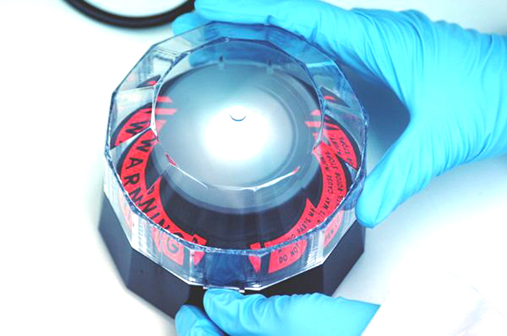
Figure 6. Drying an alkaline phosphatase microarray in a Microarray High Speed Centrifuge. Make sure the microarray is inserted completely into the plastic substrate holder before commencing centrifugation. Wear safety glasses at all times when operating this instrument.
7. Scan microarray to detect blue colorimetric signals. Use the Colorimetric Microarray Scanner to detect the blue AP signals and acquire a quantifiable TIFF image. Place one or more microarrays with the spots facing downward towards the scanner. In the proper orientation, the white membrane will be facing downwards and the corner chamfer will be in the upper left (Fig. 7). Use the SpotWareTM controller software to select scan area, resolution, gain, and other scanning parameters. A TIFF file containing the AP data is now ready for quantification and modeling.
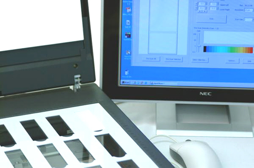
Figure 7. Developed alkaline phosphatase microarray scan-ready in a Colorimetric Microarray Scanner.
Technical Assistance
Please contact us if you have any comments, suggestions, or if you need technical assistance. By electronic mail: arrayit@arrayit.com (under the subject heading, please type ArrayIt® technical assistance). By email: arrayit@arrayit.com, Monday–Friday PST 8:30am - 5:30 PM. Please remember we want to hear about your successes!
Troubleshooting Tips
Low signal
- Increase development time
- Use SuperProtein Blocking Buffer (not BlockIt™) for SuperProtein surface
- Increase scan gain
High background
- Reduce development time
- Reduce scan gain
- Block microarray with ArrayIt® Blocking Solutions
Recommended Products
SpotBot 4 Complete Manufacturing System
Colorimetric Microarray Scanner
SuperProtein Substrates
SuperNitro Substrates
SuperEpoxy Substrates
Protein Printing Buffer
References
Click here for Arrayit Alkaline Phosphatase scientific publications.

