Pathogen Antigen Microarrays
Data Sheet
![]() Shop this product in our online store
Shop this product in our online store
Products - Protein Microarrays - Pathogen Antigen Microarrays for Rapid Detection of Serum Antibodies Against Pathogenic Microorganisms

Arrayit Pathogen Antigen Microarrays permit rapid detection of reactive serum antibodies against a wide range pathogenic microorganisms including Bartonella henselae, Borrelia burgdorferi, Brucella melitensis, Burkholderia pseudomallei, Chlamydia muridarum, Chlamydia trachomatis, Coxiella, Francisella tularensis, Mycobacterium tuberculosis, Orthopoxvirus Whole Proteome, Plasmodium falciparum, Plasmodium vivax, Salmonella enterica non-typhoidal, Salmonella enterica typhi, Staphylococcus aureus, and Toxoplasma gondii. Available as printed antigen microarrays and complete kits, this product line sets a new standard for research into the molecular basis of microbial infection.
Introduction
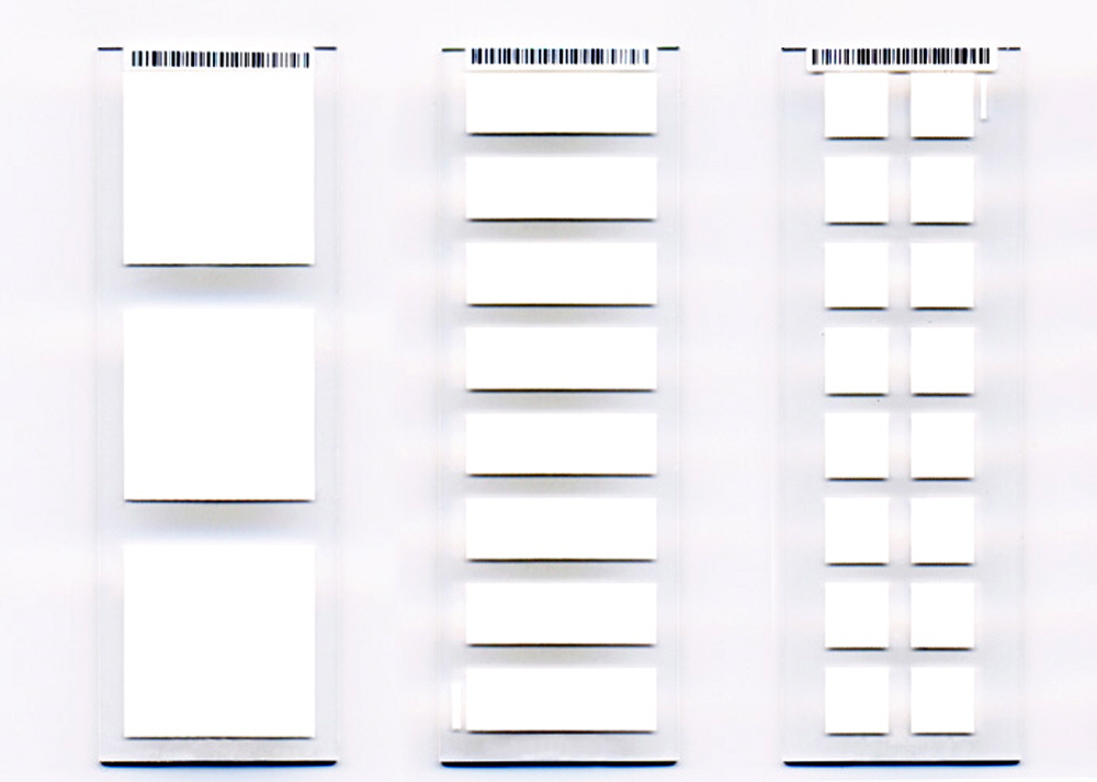
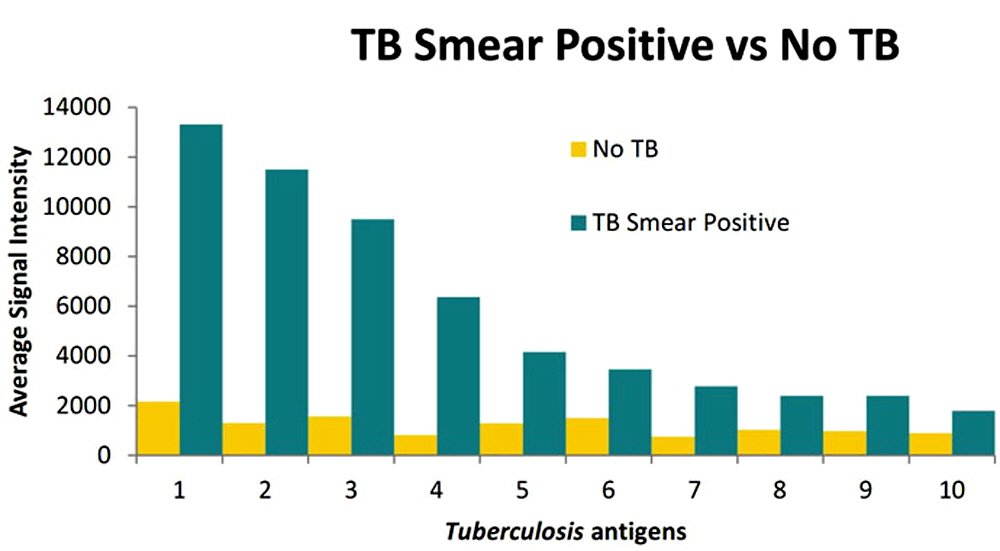
Arrayit Pathogen Antigen Microarray kits are available in 80, 160, 320 and 800 microarray packages sufficient for 80, 160, 320 and 800 serum screening assays respectively. These are highly multiplexed devices for measuring serum reactive antibodies to these pathogens. Each microarray chip contains key proteins proteins from each proteome plus an additional proteins as controls. The proteins on these microarrays are expressed by in vitro transcription/translation using an E. coli cell free expression system and printed onto nitrocellulose microarray substrate slides with each slide containing 3, 8 or 16 separate pads depending on the number of proteins for that organism. These protein microarrays have been validated by probing with serum specimen collections of disease cases and controls.

Materials supplied
- Antigen Microarrays
- Blocking Buffer
- 20X Wash Buffer
- 20X TBS
- Secondary: biotin conjugated goat anti-bovine IgG
- Tertiary: streptavidin conjugated fluorophore
- Microarray Frame, Chambers and Clips
Note: Microarray frames, chambers and clips are reusable. Additional frames, chambers and clips can be purchased from GraceBio (cat# 246870, 246853 and 204838 respectively) or other vendors who can provide equivalent assemblies.
Additional Materials Required
- Ultrapure water
- Orbital shaker or rocker platform
- Table top centrifuge
- Any microarray slide scanner with a red (Cy5) laser, available from e.g. Molecular Devices LLC, Arrayit Corporation, Agilent Technologies, and other manufacturers.
- Pipettors and tips
- Disposable polypropylene or polystyrene tubes for sample preparation
- Disposable dispensing reservoirs
- Containers for wash buffer dilution
- Timer
- Aspirator: can be assembled from a vacuum, Erlenmeyer flask, tubing, and pipette tips
Storage and Handling
- This product is shipped on cold pack or wet ice. Immediately upon receipt, store the Seconday, Tertiary, and E. coli lysate tubes at -20°C. All other components can be stored at room temperature.
- The kit is stable until the expiration date indicated on the label when stored as specified.
- The microarray slides are provided in a sealed pouch with desiccant. Once opened, any unused portion should be reclosed with desiccant in the pouch and returned to 4°C storage immediately.
- Nitrocellulose pads should not be in direct contact with any solid object, e.g. a pipette tip, as they are susceptible to scratching.
- Allow reagents to warm up to room temperature before use.
- Do not use any reagent beyond the expiration date.
- The diluted 1X Wash Buffer is stable the day of the preparation only.
- Avoid exposure of fluorophore to light, air, or extreme temperatures.
Precautions
- Read the manual before starting work.
- Human serum is potentially pathogenic and should be handled in a class II biological containment hood. Wear appropriate PPE (disposable gloves, lab coat) when handling serum. Avoid aerosols and remove sharps from the work area. All liquid waste containing human serum should be inactivated with 10% (final concentration) chlorine bleach or equivalent.
- All kit reagents should be handled according to good laboratory practice. Wear appropriate PPE (disposable gloves, lab coat). Use a dedicated workbench. No eating, drinking, applying cosmetics, smoking etc in the area. Follow laboratory guidelines for liquid and solid waste disposal or consult your biological safety officer.
Specimen collection and preparation
- Collect serum or plasma specimens. Serum should be separated from red blood cells as soon as possible.
- If storing specimens, avoid repeated freezing and thawing.
Reagent Preparation
- Reconstitute each lyophilized E.coli lysate (ECL) tube with 1.2 mL blocking buffer. Surplus lysate can be stored at -20°C.
- Dilute wash buffer and TBS to 1X concentration. Diluted buffers are only stable the day of preparation. Be sure to only dilute the amount of buffer needed.
Procedure
DAY 1:
- If frozen, thaw serum samples on ice
- Assemble the microarray frame, chamber and slide. Cover with a lid. Assembly of a 8-pad slide is shown below; the 3-pad slide contained in this kit is assembled in an identical manner.
Figure 1. Frame, Chamber, clips and 8-pad slide before assembly
- Figure 2. Align the 8-pad slide and the chamber
- Figure 3. Snap one clip over one edge of the 8-pad slide/chamber
- Figure 4. Snap a second clip over the other edge of the 8-pad slide/chamber
- Figure 5. Place the completed 8-pad slide/chamber/clip into the frame
- Figure 6. Finished assembly (up to 3 slides can be assembled into the frame)
- Block the microarrays with Provided Blocking Buffer per well for 30-60 minutes.
- Make 10% ECL in Blocking Buffer for pre-incubation of serum samples. Now referred to as pre-incubation solution.
- In a Biosafety Cabinet, dilute serum 1 to 100 in pre-incubation solution and incubate for at least 30 minutes (350 µl/well needed for a 3-pad).
- In a Biosafety Cabinet, remove blocking buffer from pads and immediately add diluted serum samples. Do not let the pads dry out. Put the frames into a humidified airtight container. Incubate overnight at 4 degrees with gentle agitation.
DAY 2:
- Wash with 1mL 1X wash buffer per well 3 times for 5 minutes each. Wash by aspirating the serum solution and immediately adding 1 mL 1X wash buffer, being careful not to let the pads dry out.
- Make 1% ECL in Blocking Buffer for dilution of secondary antibody (Biotin conjugated goat anti-bovine IgG Fcγ) and Streptavidin conjugated fluor. Now referred to as blocking solution.
- Dilute secondary antibody 1 to 1,000 in blocking solution (350 µl/well needed for a 3-pad).
- Remove wash buffer and add 350 µl diluted secondary antibody per well.
- Incubate for 1 hour at room temperature.
- Wash with 1 mL 1X wash buffer per well 3 times for 5 minutes each.
- Dilute tertiary detection fluor 1 to 200 in blocking solution (350 µl/well needed for a 3-pad). Protect from light until ready to use.
- Remove wash buffer and add 350 µl diluted tertiary detection fluor per well.
- Incubate for 1 hour at room temperature in the dark (i.e. cover with aluminum foil).
- Wash with 1 mL 1X wash buffer per well 3 times for 5 minutes each.
- Wash with 1 mL 1X TBS per well 3 times for 1 minute each.
- Fill a 2L beaker with ultra pure H2O.
- Remove slide from Fast frame assembly and dip in ultra pure water. Place into a 50 ml conical with a Kimwipe at the bottom. Spin at room temperature for 5 minutes at 500X g to dry.
- Scan slides, quantify and analyze data.
- *Note: All incubation and washes are done on a rocking platform. First washes after incubation should be more thorough, using an aspirator, but subsequent washes may discard solution by dumping as long as wash buffer can be added quickly to prevent drying of the pads.
Quality control
Each microarray contains positive and negative control spots.

Data Analysis
- Customers may choose their desired methods and software to perform data analysis.
- If desired, data analysis service may be arranged with ADi upon request.

Alternative Detection Methods
- The Biotin-Streptavidin fluorophore detection system provided in this kit has demonstrated the best sensitivity throughout our extensive research and development. However, customers may choose alternative procedures to detect the reactive antibodies in the human serum sample to suit their specific needs. These include alkaline phosphatase conjugated secondary antibody with a coloarimetric detection system and fluorophore conjugated secondary antibody.
- Furthermore, customers may detect the reactive antibodies in serum obtained from other animals by substituting a secondary antibody against the subject animal. For instance, if the customer is interested in detecting antibody in bovine serum samples, biotin conjugated goat anti-bovine IgG Fcr (which can be purchased from many sources) would be substituted for the human secondary antibody contained in this kit.

